
Bone Changes: Reproductive Avian Female
Polyostotic Hyperostosis
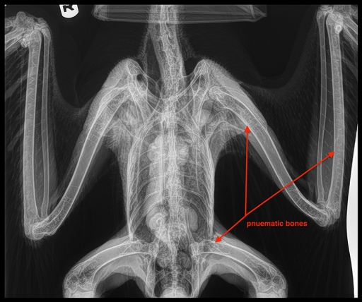 Bird bones are unique in their structure and function. Many of the bones are considered pneumatic: bones that contain small diverticula (sac or pouch) originating from the air sacs. These diverticula, filled with air, give the bone marrow a corrugated appearance versus a solid appearance of a non- pneumatic bone (fig 1). Examples of pneumatic bones include the skull, clavicle, keel, pelvic girdle, lumbar and sacral vertebrae, and the long bones (femur, tibia, humerus).

Reproductively active female birds will often show bone changes in the radiographs of the long bones (femur, tibia, humerus), pelvic girdle, and vertebrae. This is a normal change referred to as polyostotic hyperostosis. Radiographic evaluation of a polyostotic bone will show an increase of bone density, especially the long bones.
Medullary Bone Development
The cavity of the bone is where red bone marrow and yellow bone marrow (adipose tissue) is produced and stored. The common name is the marrow cavity. Medullary bone is considered a labile bone, made of cells that multiply constantly throughout life. In normal circumstances, medullary bone occurs only in the female bird during the reproductive phase. Medullary bone development is under the influence of the ovarian hormones: estrogen and testosterone.

 In the long bones, as medullary bone develops, a system of interconnecting spicules grow from the endosteal surface of the cortical bone (dense outer surface of the bone) and penetrate into the marrow cavity. Calcium is stored in the bones during this time and will change the appearance of the pneumatic bone to a somewhat solid mineral dense bone (fig2). A complex association of endocrine hormones helps to regulate the deposition of calcium and formation of this medullary bone. Research in the chicken has shown that medullary bone formation in the female occurs during the final 10 days before egg laying.


Research has also identified that intense medullary bone formation alternating with periods of severe bone depletion takes place during the ovulation-oviposition cycle and shell calcification. Birds on a proper diet with adequate calcium are generally able, during the time when shell production is not active, to replenish the calcium lost from the medullary bone during shell calcification. Once the estrogen levels return to normal and the female is no longer reproductive, the bone densities usually return to normal. Once again the bones will show the typical pneumatic pattern. These are normal changes in the reproductively active female, as this bone is the primary source of calcium for egg shell formation.
Pathologic Development of Medullary Bone
 Polyostotic hyperostosis is found in varying degrees in the female of many species and simply represents the normal hormonal changes associated with egg formation. However, there are several situations where abnormal development of this condition may present itself. During reproductive activity, a diet deficient in calcium and Vitamin D3 may result in cortical bone being eroded as the body tries to maintain the proper formation of medullary bone. This will lead to only partially calcified medullary bone and a moth-eaten appearance to the bones on radiographs. Certainly, proper egg shell formation can be negatively affected if the medullary bone formation is inadequate. Female birds suffering from hyper-estrogenic conditions, such as cystic ovarian disease or ovarian neoplasia, will show forms of prolonged polyostotic hyperostosis (fig 3).

Polyostotic hyperostosis can also occur in male birds. This is typically associated with the development of testicular tumors, most notably a sertoli cell tumor that produces estrogen.
Treatment for Pathologic Development of Polyostotic Hyperostosis
As mentioned above, the female bird with normal reproductive activity will not need any intervention for this condition. This is a normal change in the bones with egg shell formation. Female or male birds with reproductive- associated disease will require medical intervention. Radiographs will evaluate the skeleton looking for changes supporting polyostotic hyperostosis, and evaluate organ enlargement and displacement, while chemistry profiles will evaluate organ function. Tumors of the reproductive tract often require surgery – with removal of the tumor, orchiectomy (removal of a testicle) or salpingectomy (removal of the oviduct). The prognosis for long-term survival is dependent on the type of neoplasia present and the possibility of metastatic disease.
Some reproductive diseases will respond to conservative treatment with GnRH-agonists that suppress reproductive activity in the bird. Lueprolide acetate and Deslorelin acetate implants are two common medications used to treat reproductive-associated disease in both males and females. Lueprolide acetate is given as an intramuscular injection and may require multiple injections to achieve treatment of the reproductive disorder. Deslorelin acetate is an implant, usually administered intramuscular, providing therapeutic levels for up to 3 months or longer, depending on the species of bird and reproductive disease present. In male birds with a sertoli cell testicular tumor, these medications may help counter the effects of estrogen produced by this tumor, enhance and maintain quality of life.
Suggested Reading